Deprivation amblyopia: when the image is blocked from the start
Published: July 27, 2026
In short
In deprivation amblyopia the brain never receives a proper image from one eye, so development stalls from the outset. It is the rarest form but the one needing the fastest response, and removing the obstacle is usually not enough on its own.
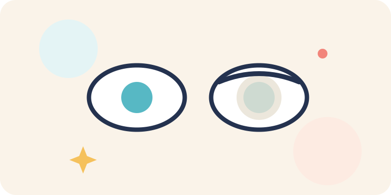
The two most common causes of lazy eye are a difference in prescription and strabismus. There is a third route: the image never reaching the retina at all. This is deprivation amblyopia, the rarest form and the most urgent.
This article covers how the obstruction arises, why it is a race against time, and why removing the obstacle is not enough by itself.
How is the image blocked?
The best-known cause is a congenital cataract: the lens of the eye is cloudy and does not let light through cleanly. The result is that the eye never sends a sharp image to the brain.
The second common cause is a drooping eyelid (ptosis), where the lid covers the line of sight partly or completely. More rarely, a clouding of the cornea or another obstruction inside the eye produces the same outcome.
The common thread is this: the problem is not in the brain comparing two images but in one image never forming at all.
Why is it so urgent?
The visual system is built rapidly in the first months and years, and that construction depends on regular input from the eye. If the input never arrives, the relevant connections never get their chance to develop.
In other forms of amblyopia the brain still receives an image, blurred though it may be; in deprivation the signal is almost entirely absent. The effect is deeper and the time pressure higher.
This is among the main reasons infant eye checks are done without waiting for any complaint.
Does removing the obstacle end the problem?
No, and this is where families are most often surprised. Removing a cataract or correcting a lid clears the path; the brain learning to use that eye is a separate process.
After surgery, appropriate optical correction and then vision training are usually planned. Surgery is the beginning, not the end.
The order and the method are set by the doctor according to the child's age and the details of the picture.
Signs a family might notice
A whitish appearance in a baby's pupil, one eyelid sitting markedly lower than the other, or one eye responding noticeably less to light or to faces are signs worth attention.
None of them is a diagnosis on its own, but noticing any of them is reason enough to see an eye doctor without waiting.
Terms used here
- Deprivation amblyopia
- Lazy eye that develops because the image is physically prevented from reaching the retina.
- Congenital cataract
- A clouding of the lens present from birth, blocking light from passing through.
- Ptosis
- A drooping upper eyelid, which can cover part of the field of vision.